In a paper published in the journal Science, the NU researchers explain that they are taking a first step towards addressing this problem by focusing on the enzyme called particulate methane monooxygenase (pMMO), which the bacteria use to catalyze the reaction. The substance, however, is a particularly difficult protein to study because it’s embedded in the cell membrane of the bacteria.
Typically, when researchers study these methanotrophic bacteria, they use a harsh process in which the proteins are ripped out of the cell membranes using a detergent solution. While this procedure effectively isolates the enzyme, it also kills all enzyme activity and limits how much information researchers can gather.
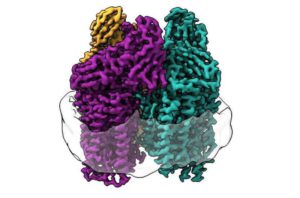
To avoid this issue, the scientists put the enzyme back into a membrane that resembles its native environment. They used lipids from the bacteria to form a membrane within a protective particle called a nanodisc and then embedded the enzyme into that membrane.
“By recreating the enzyme’s native environment within the nanodisc, we were able to restore activity to the enzyme,” Christopher Koo, first author of the study, said in a media statement. “Then, we were able to use structural techniques to determine at the atomic level how the lipid bilayer restored activity. In doing so, we discovered the full arrangement of the copper site in the enzyme where methane oxidation likely occurs.”
The scientist and his colleagues used cryo-electron microscopy, which allowed them to visualize the atomic structure of the active enzyme at high resolution for the first time.
“As a consequence of the recent ‘resolution revolution’ in cryo-EM, we were able to see the structure in atomic detail,” senior author Amy Rosenzweig said. “What we saw completely changed the way we were thinking about the active site of this enzyme.”
Rosenzweig said that the cryo-EM structures provide a new starting point to answer the questions that continue to pile on. How does methane travel to the enzyme active site? Or does methanol travel out of the enzyme? How does the copper in the active site do the chemical reaction?
Given these questions, the team’s next step is to study the enzyme directly within the bacterial cell using a forefront imaging technique called cryo-electron tomography.
If successful, they will be able to see exactly how the enzyme is arranged in the cell membrane, determine how it operates in its truly native environment and learn whether other proteins around the enzyme interact with it. These discoveries would provide a key missing link to engineers.
“If you want to optimize the enzyme to plug it into biomanufacturing pathways or to consume pollutants other than methane, then we need to know what it looks like in its native environment and where the methane binds,” Rosenzweig said. “You could use bacteria with an engineered enzyme to harvest methane from fracking sites or to clean up oil spills.”


